Contact numbers: 598190806/588883230
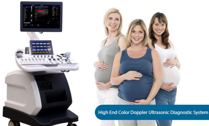
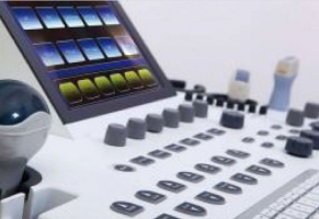
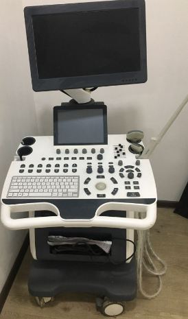
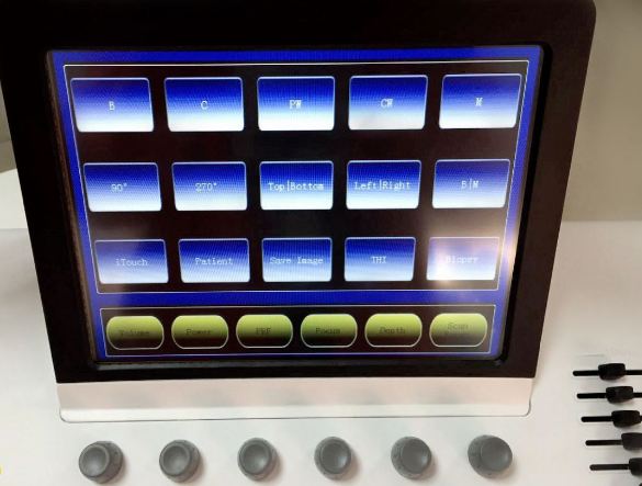
Display: 19 liquid crystal display, 10.4 touchscreen.
Power supply: power alternating current (220 volts + -22 volts, 50 Hz, + - 1 Hz
Input power: 350 volts
Electronic shock protection: Class I
Quality of Electronic Shock Protection: BF Type
Protection against damaging water entry
IPX1 probe cable: IPX1
IPX7 probe trans door: IPX7
Scan mode
Electronic: linear, convex, phased grid
Screen type:
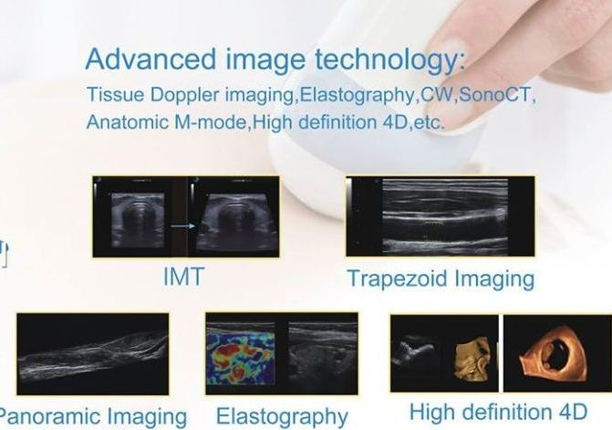
Black-and-white image: B, 2B, 4B, right / left B / M, B / D Right PW (D), Upper / Lower PW (D), M, B, Local mode amplification, linear probe trapezoidal visualization, 3D Reconstruction.
Blood flow images: Spectrum: B / BC, BC / B, B / C / D, C / F / M, B / C / D, B / C / M, B / C Double Real Time, PW (D) ( D), CFM, CPA
Color Doppler
Screen type: energy, speed, variation (to help speed variation), frequency, amplification.
Image control: (Sample selector) Sampler size and position adjustable, linear probe radiation angle adjustable.
Spectral Doppler: Sampler volume and position adjustable.
PW (D) search has maximum and medium speed support,
Max. The speed limit is adjustable
Scanner Field: Heavy above the baseline, below the baseline and full scan.
PW (D) Sound on / off and height adjustment.
Speed measurement: Maximum / minimum speed support
Blood flow image control options: frequency, sampler position and size, base line, color enhancement, turning angle heavy, wall filter, cumulative amount.
Image color intensity: constantly adjustable
Focus: Electronic focus + sound lens focus
Radiation includes, 1/2/3/4 foci.
Continuous dynamic focus when receiving: Digital beam formation
Continuous dynamic focus
Dynamic groove diameter
The weighted sum of the delay in receipt
Scanning SEMI-MOMENT and supporting + - 10-degree linear mile turning angle,
Multi-beam processing
Signal Percentage / Doppler Sound
Dynamic wave filtering
Square demodulation
Total reinforcement control
Composite reinforcement technology:
8 Sexual TGC and D-AGC
Low frequency filter.
Stereophone acoustic edition of adjustable Doppler adjustable acoustic output data
Image processing Dynamic range variation and logarithmic complexation,
The lung of time
Spatial filtering
Shot medium
Expansion of the trunk
Gray scale variation
Scanning speed in B / M or M mode, linear density control
Scan angle / width control
Image optimization
Switch the image up and down.
Multi-angle rotation of the image
Choice of color gray-scale barrier inversion
Combined processing of fabric and power
Resolution of black and white images: 1024 X 778 X 8 bits (256 gray - scale)
Color image resolution: 8 bits x 8 bits x 8 bits x color coding
Choose specific images
Optimized items are available for selection
Basic measurement and computational functions
2D Measurement: Distance, Angle, Perimeter, Area (Ellipse and Locus Models)
Volume - three-line method (length-width-height)
Histogram, transverse - crossing scheme,
Basic MM - Rhythm Measurement: Time, Distance, Heart Rate, Valve Frequency
Childbirth measurement
4 types of database to calculate the duration of childbirth
3 irreplaceable versions: Asian, European, American, + 1 interchangeable version, individualized for customers
The duration of childbirth can be calculated in all versions: the diameter of the fetal membrane, the size of the fetus before the start, the biparietal diameter, the circumference of the head, the circumference of the abdomen, the length of the thigh, the length of the shoulder bone, the diameter of the abdomen, the size of the spine, the diameter of the forehead The length of the bone, the length of the radial bone, the cistern magnum, the inside diameter of the waltz nest, the diameter of the eye socket, the outside diameter of the eye socket, the amniotic fluid index, the fetal heart rate, the transparency of the occiput, the distance and area, and so on. Sh.
Childbirth report
Amniotic fluid index measurement and calculation (AFI)
Area Calculation (BPD / OFD, FL / AC, FL / BPD and HC / AC)
Fetal Weight Assessment (EDDD) Calculation with LMP and BBT, Fetal Biophysical Assessment, Fetal Growth Diagram, Andrology Measurement and Calculation, Prostate and Testicular Prostate Antifungal Calculation, Calculation of Expected Prostate Calculation of density (PSAD)
Gynecological measurement: measurement and calculation of the uterus, left ovary, right ovary, left and right follicles
Urological measurement and calculation: Diagnosis and measurement of left ventricular, bladder, residual urine volume.
Vascular Measurement and Calculation: Measurement / Calculation of Area Stenosis Rate and Vascular Stenosis Rate
Diagnosis and measurement of multiple fetuses, diagnosis and calculation of small inclusions
Diagnosis and calculation of thyroid, galactophores, masses, etc.
Cardiac diagnostics and calculus functions
The cardiology measurement software package includes: aortic, mitral valve, left / right ventricular analysis and measurement capabilities.
Percentage of area stenosis (% area stenosis), tube stenosis diameter (% diam. Sten)
Body Surface Area (BSA): Body marking database is divided into several parts according to body function: abdominal cavity, andrology, cardiology, gynecology, bone muscle, obstetrics, pediatrics, small parts, urology, and so on. Sh
Information displayed on the screen: probe conditions, image type, depth, focus, dynamic range, body marking, probe position, voice output, patient status, medical agency name, measured size, time and number, scale, scan direction, gray scale diagram . The current frequency of the probe, the frame rate, the increase in the b-c-d mode as a whole.
Menu: Annotation Gray Scale Zone Function Line, TI (Thermal Index and MI Mechanical Index)
Memory (warehouse):
Probe setting
Image
Apparatus cycle
Measurement results
Report
Automatic coverage of the apparatus cycle, manual coverage, adjusting the speed of the cycle, searching for the apparatus cycle (front-to-back), front / rear coverage.
256 Gray Scale
In bilingual interface mode: Chinese and English
The function of the small ball to move the cursor, the performance of various operations depends on the job status and menu. By controlling several electronic scales, determining the cursor, location, completing the measurement.
Entrance - exit port
VGA - DVI
Network port
Video port
USB port
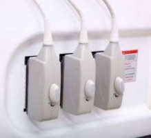
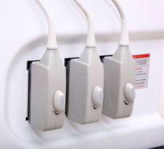
Probe: 3.5 mm hertz, R 65 Multi-frequency convex probe with 128 channels,
The frequency range of the probes is: between 2.0 miles and 5.5 miles.
The scanning angle in B mode is 140-158 degrees.
M mode can activate specific matrix element as well as unchanged frequency probes.
Can be used in combination with B-M mode
Scope of use:
Tasting and diagnosis of abdominal tissue such as: liver, gallbladder, spleen pancreas, kidney, gastrointestinal tract, uterus, bladder, prostate, etc. Sh.
Evaluate fetal maturation and growth at the time of diagnosis
6.5 Mile Hertz R10 Multi-Conducting convex probe (with 128 channel)
Areas of application: vagina, uterus, rectum, rectal wall, prostate and so on.
7.5 Mil Hertz, L 46 Multi-frequency, linear probe
(128 channels)
The frequency range of the probes is: 6.0 miles between Herz and 12.0 Mil Herz.
The scanning width in B mode is 38-45 degrees.
Areas of application: Newborns, musculoskeletal system. Peripheral blood vessels, small inclusions, including mammary, thyroid, and testicular glands.


